Most Common Misdiagnosed Ligament Injuries in the Neck (AKA) Cervical Instability and Injury.

Did you know…. the TOP 30% of the cervical spine is not properly analyzed by non weight bearing motionless static X-rays, MRIs, or CT scans?

Most Common Misdiagnosed Ligament Injuries in the Neck (AKA) Cervical Instability and Injury – innovative diagnostic technology
This is the area of the cervical spine between the skull (C0), 1st cervical vertebrae call atlas (C1),
and the 2nd vertebrae called axis (C2).
Did you know….there are NO spinal discs and only ligaments between the skull (C0), 1st cervical vertebrae called atlas (C1), and the 2nd vertebrae called axis (C2)?
These small ligaments stabilize and hold the upper cervical spinal joints together preventing excessive motion between the skull (C0), 1st cervical vertebrae atlas (C1),and the 2nd vertebrae axis (C2).
Did you know….. the joints that make up the top 30% of the cervical spine between the skull (C0), 1st cervical vertebrae call atlas (C1),
and the 2nd vertebrae called axis (C2) are responsible for 50% of the cervical flexion and rotation?
The function of a cervical ligament is to stabilize and prevent excessive motion between the spinal bones when we move. If there is excessive motion between the spinal bones this indicates ligament damage, injury, and laxity.

Most Common Misdiagnosed Ligament Injuries in the Neck (AKA) Cervical Instability and Injury – innovative diagnostic technology
Did you know….. you can not properly test the function of a cervical ligament without movement of the spinal joint?
What is the best weight bearing motion based study to analyze the 22 major ligaments in the spine?
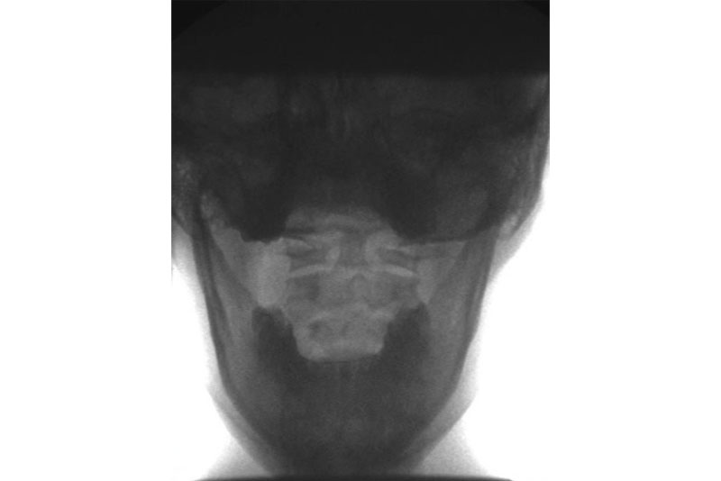
DMX (Digital Motion X-ray) is an image-intensified fluoroscopic x-ray system that allows the doctor or clinician to perform weight bearing x-rays on a patient while moving the joint or body part.
The digital motion x-ray makes a digital video of the actual joint or body part in motion (spine, hip, knee, ankle, foot, shoulder, elbow, wrist, hand, and TMJ).
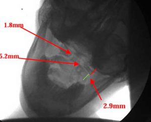
DMX motion x-rays are analyzed with Computerized Radiographic Mensuration Analysis (CRMA) to achieve accurate measures and to determine the extent of the injury.
DMX is an innovative diagnostic technology that can help diagnose painful conditions that are often undetectable by motionless static x-rays, MRIs, and/or CT Scans, MRI’s (magnetic resonance imaging).
Common Symptoms Of Cervical Ligament Damage?
Headaches/Migraines
Dizziness
Unstable Feeling
Blurred vision
Brain Fog
Difficult Concentrating
Pain increased with movement
Localized neck pain
Pain the the traps and/upper mid back
Pain radiating into the head and eyes
Pain radiating into shoulders arms hands and/or fingers
Numbness in the shoulders arms hands and/or fingers
Weakness in the shoulders, arms, and/or hands
If you have been experiencing spinal symptoms such as neck pain, back pain, headaches, migraines, mid back pain, rib pain, carpal tunnel, sciatica, disc herniations or protrusions, neuropathy, scoliosis, spinal stenosis, pain/numbness/weakness in your arms, hands, legs or feet……..
Call our office Today (305) 275-7474 for a FREE Phone, Whatsapp, or Zoom consultation so we can determine the cause of your symptoms, identify the damage, and design an individualized treatment plan to get back to enjoying the thing you love doing!
https://sunsetchiropractor.com/appointment/
We look forward to meeting you and helping you recover your health. You can get better! We can help! There is still hope !
Dr. Rodolfo Alfonso, D.C.
Dr. Mark N. Berry, D.C.
Sunset Chiropractic and Wellness
8585 Sunset Dr.
STE 102
Miami, Florida 33143
305-275-7474
www.sunsetchiropractor.com
[button size=”big_large_full_width” icon=”fa-medkit” target=”_self” hover_type=”default” text_align=”center” text=”Corrective Chiropractic Care” link=”https://sunsetchiropractor.com/corrective-chiropractic-care/” color=”#ffffff” background_color=”#9b7f60″ icon_color=”#ffffff” hover_border_color=”#ffffff”]
Did you know….. you can not properly test the function of a cervical ligament without movement of the spinal joint?
What is the best weight bearing motion based study to analyze the 22 major ligaments in the spine?
DMX (Digital Motion X-ray) is an image-intensified fluoroscopic x-ray system that allows the doctor or clinician to perform weight bearing x-rays on a patient while moving the joint or body part.
The digital motion x-ray makes a digital video of the actual joint or body part in motion (spine, hip, knee, ankle, foot, shoulder, elbow, wrist, hand, and TMJ).


